Skip to main content
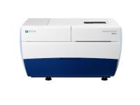
This high content, high throughput confocal imager is a high speed, automated, confocal imager which contains live cell imaging and 7 channel florescence laser excitations. It also includes high-end data analysis modules for deep learning segmentation and phenotype classification. The system is directly relevant to cancer research and regenerative medicine research, especially for drug discovery, translational and clinical research.
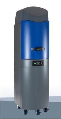
EXODUS is an automatic, label-free, and highly efficient exosome isolation system. With EXODUS, you can easily and quickly isolate high-quality, intact exosomes with excellent yield and purity from a variety of bio-fluids and sample volumes. EXODUS has been developed using a dual-membrane nanofiltration system that integrates periodic negative pressure oscillation (NPO) and double-coupled ultrasonic harmonic oscillations (HO).
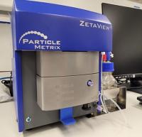
Nanoparticle tracking analysis (NTA) visualizes particles in liquid suspension using laser illumination and utilizes an ultramicroscope to analyze particle number, size, and zeta potential. This allows for measurement of particles 10-1000 nm in diameter, including extracellular vesicles (EV). In our laboratory, the ZetaView Particle Metrix performs NTA as part of our EV characterization workflow.
Size exclusion chromatography (SEC) separates particles based on their size by utilizing the differences in their flow rates through a column filtering system. As samples flow through beads in the column, they are separated in order of decreasing molecular weight. In our research, we use an Izon Automated Fraction Collector for SEC to both isolate and purify EVs.
Tangential flow filtration (TFF) is a rapid and scalable method of isolating and purifying cells, extracellular vesicles (EV), and biomolecules. The system utilizes a peristaltic pump to push sample fluid through a filter with the fluid flow parallel to the hollow fiber membrane pores. In our group, TFF is being explored as a more efficient alternative to the gold standard ultracentrifugation method for EV isolation.
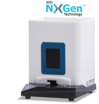
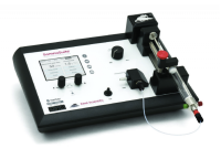
The NanoAssemblr® Spark is part of the NanoAssemblr® platform from PRECISION Nanosystems. It is the only microfluidic system designed and engineered to produce nanoparticles for all stages of nanomedicine development. The Spark has been developed to make ultra-low volume nanoparticle production efficient and reproducible with unmatched ease and simplicity in one step process.
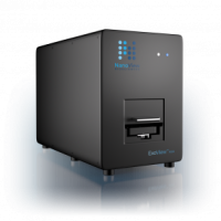
ExoView R100 is a fully automated platform that provides multi-level and comprehensive measurements for extracellular vesicle (EV) particle size analysis, EV count, EV phenotype, and biomarker colocalization. The ExoView platform provides previously unattainable information in a single and bias-free sample workflow. ExoView works without the need for sample purification and brings the researcher one step closer to analyzing a sample in its natural state. ExoView R100 is an affinity-based technology that allows specific populations of exosomes and other extracellular vesicles to bind in a multiplexed manner to a functional ExoView chip. The fully automated instrument can measure 9 samples automatically, direct from the sample, saving cost, time, and reducing purification biases.
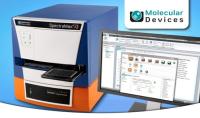
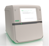
The SpectraMax® i3 Platform from Molecular Devices® is a multi-mode detection system that can read plates using standard spectral absorbance, fluorescence, and luminescence detection. We use the SpectraMax MiniMax to image cells and analyze them with SoftMax Pro Software.
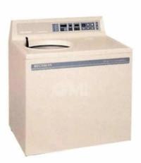
Designed to eliminate a high-speed vacuum seal, the L7 Ultracentrifuge carries a vacuum-encased induction drive which reduces heat and noise output. In our research, we use the SW28 rotor for EV isolation.
The Lago X Optical Imaging System provides a powerful and flexible in vivo imaging capability by delivering an unmatched 10 mouse capacity across BLI, FLI and X-ray. The imaging system contains a high-performance cooled CCD camera to record the image collected by a large aperture lens with automation for filter and field of view selection. The combined luminescence, fluorescence, and X-ray modalities of this system enable us to conduct high throughput imaging for our animal studies.
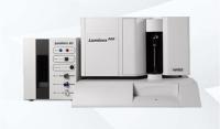
The Luminex platform comprises Luminex instruments and multiplex assays for measuring RNA and protein targets. Luminex multiplex assays use xMAP bead-based technology to simultaneously detect and quantitate multiple secreted proteins including cytokines, chemokines, and growth factors. This multiplex technology can also be used to measure gene expression by utilizing the Invitrogen QuantiGene Plex assays. The Luminex system produces results comparable to conventional assays such as ELISA and qPCR, but with greater efficiency and throughput. The Luminex 200 Instrument System sets the standard for multiplexing, offering us the ability to perform up to 100 different tests in a single reaction volume on a flow cytometry–based platform.
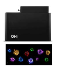
Super-resolution microscopy surpasses the resolution barrier of conventional light microscopes by enabling detection and quantification of single proteins and nucleic acids at the sub-vesicular level. For our research, we use the Oni Super Resolution Microscope to reconstruct vesicle membrane composition with single-molecule localization microscopy (SMLM), to identify the specific biomolecules involved in EV signaling and targeting, and to identify and visualize sub-populations of EVs. We are currently looking into how this data may provide important information about the role EVs play in disease progression.
This instrument is designed to perform numerous tests on tissues and other biomaterials including: tensile, compression, flex, peel, puncture, friction, and shear tests. In our laboratory, we use the Instron Mechanical Tester to analyze the strength of tissues and our bioengineered scaffolds.
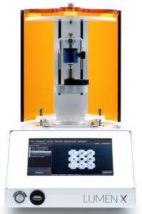
The Lumen X is a digital light projection (DLP)-based 3D bioprinter capable of printing multi-vascular and functional intravascular networks for biomedical applications. With feature resolution of 50 µm and compatibility with versatile materials, the Lumen X enables high resolution and consistent printing. This is one of the instruments we use to print components of our bioengineered scaffolds.
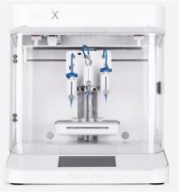
Bio X 3D Bioprinter is an extrusion-based bioprinting system with a printhead temperature range of 4°C to 250°C and a printbed temperature range between 4°C and 65°C. This design allows us to utilize a wide range of materials and cell types to construct our bioengineered scaffolds.
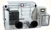
Hypoxic Chamber
O2 Control Glove Boxes are equipped with a purge-only airlock, which is a transfer chamber that equilibrates O2 levels by purging excess O2 prior to opening the door into the actual glove box and placing items inside. The Humidified Incubation Box for Cell and Tissue Culture is a separate unit that sits inside the glove box which allows our cultures to be humidified with the same atmosphere content (gas and temperature) without immediately increasing the humidity of the rest of the box.
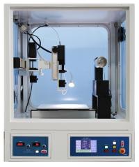
Electrospinning System
Electrospinning systems can reconstruct organic and inorganic material solutions into nanofibers through high voltage. These instruments are widely used in our laboratory to manufacture nanofibours scaffolds for tissue engineering applications.
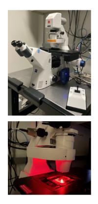
This is an advanced inverted fluorescence microscope with a motorized stage, two AxioCam cameras (MRc for color and MRm for fluorescence), and a new illumination system (Lumen 200; PRIOR) with a long lasting 200-watt metal arc lamp known for its extended transmission in UV and IR. There are also four fluorescence filter sets: DAPI, GFP, Texas Red and Cy5 for multi-fluorescence imaging. Additionally, there is an Aptome 3 attachment, which allows for optical sectioning of fluorescent samples and eliminates out of focus light. We use this microscope with Zeiss Zen One software for many different cell imaging protocols.
SomnoSuite low-flow anesthesia delivery system designed specifically for mice and rats. This is engineered with a precision syringe pump and integrated digital vaporizer which uses either room air or compressed gas to deliver anesthesia at low flow rates proportionate to the animal's size.
Small Animal Surgical Suite
Our small animal surgical suites contain two dual head operating microscopes with a focal range of 0.7 - 4X magnification. Dual head orientation allows for surgeons to operate simultaneously on an animal and dual gooseneck high powered LED lights provide illumination while working. The surgical suites are also equipped with two low profile Kent Scientific SomnoSuite Isoflurane anesthesia machines and temperature control heating pads to maintain normothermic body temperature during surgical procedures.
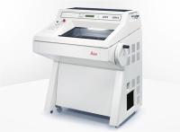
The Leica CM3050 S delivers high-quality tissue sections rapidly and efficiently by utilizing a quick-freezing shelf with defrost function and a powerful refrigeration system. The precise specimen orientation and the specimen feed system guarantees reproducible, thin, serial sections of tissue that we use routinely for histological purposes.
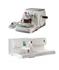
The Autocut automated microtome allows for fast and precise sectioning of histology samples. The Arcadia modular tissue embedding system incorporates an Arcadia H heated embedding workstation and an Arcadia C cold plate which offers us the flexibility to arrange the embedding workflow in any direction. The cold plate is equipped with an environment adaptive control module to ensure our samples are stabilized at -6°C.
CosMx SMI is the first high-plex in situ analysis platform to provide spatial multiomics with formalin-fixed paraffin-embedded (FFPE) and fresh frozen (FF) tissue samples at cellular and subcellular resolution. CosMx SMI enables rapid quantification and visualization of up to 1,000 RNA and 100 validated protein analytes. We utilize this spatial single-cell imaging platform for cell atlasing protocols, tissue phenotyping, cell-cell interaction imaging, cellular process imaging, and biomarker discovery.
The ChemiDoc Imaging System delivers fast, reliable, and sensitive imaging of gels and chemiluminescence western blots. This system is capable of imaging with stain-free technology, chemiluminescence detection, and a wide range of gel stains. In our research, we use the ChemiDoc Imaging System to document gels and western blots for quantitative analysis.
The RWD Spinal Impactor is an instrument utilized in injured spinal cord models to simulate the biomechanical characteristics of trauma. It is specially designed to assess mechanisms of the spinal cord model and provides valid, uniform and repeatable results through a software interface. We utilize this instrument in particular for our spinal cord injury studies for its reliability and ability to solve modeling error issues related to spinal collapse.
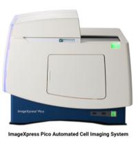
The ImageXpress® Pico Automated Cell Imaging System is a high-resolution digital microscope capable of fluorescence imaging or brightfield assays. This instrument utilizes CellReporterXpress® Image Acquisition and Analysis Software to allow for preconfigured protocols for a multitude of cell-based assays to accomplish rapid imaging in a user-friendly manner. We use this in our research to image stained tissue for histological analysis.
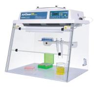
AirClean® Systems AC600 Series PCR Workstations prevent cross-contamination during DNA and RNA amplification by utilizing ISO 5 HEPA-filtered air and UV light irradiation. This system creates a clean space in our lab for DNA and RNA work.
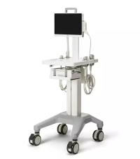
Philips InnoSight Compact Ultrasound System with doppler allows for convenient, accurate and mobile ultrasound imaging even in clinical settings. We utilize this instrument for transabdominal imaging of fetuses for surgical precision and diagnosis in our animal studies.
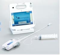
The Mcompass Anorectal Manometer is the first portable device for anorectal manometry. It can be utilized for many purposes, including diagnosis of trauma-related conditions. We use this instrument in our research to access functional outcomes from MMC treatments in our larger sheep studies.
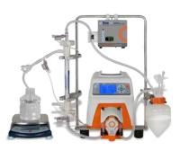
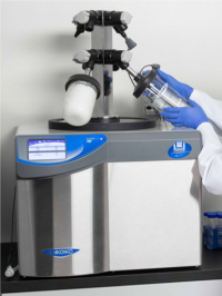
FreeZone Freeze Dryer from LABCONCO is designed to handle the lyophilization needs of research and pilot plant laboratories. All models include a large touch screen display. The system provides Real-Time Sample Data and Consistent, High Quality Results for lyophilization.